Light Microscopy Core
Hours
Monday through Friday, 8 a.m. to 4:30 p.m.
Users who have received training and are wanting to use the Light Microscopy Core after hours can obtain access from Sarah Abrahamson. Policies regarding the use of the facility after hours are listed on the Policies page.
Location
Suite W162 on the first floor of the UND School of Medicine and Health Sciences building, 1301 N. Columbia Road, Grand Forks.
Instrumentation
Confocal Microscope Systems
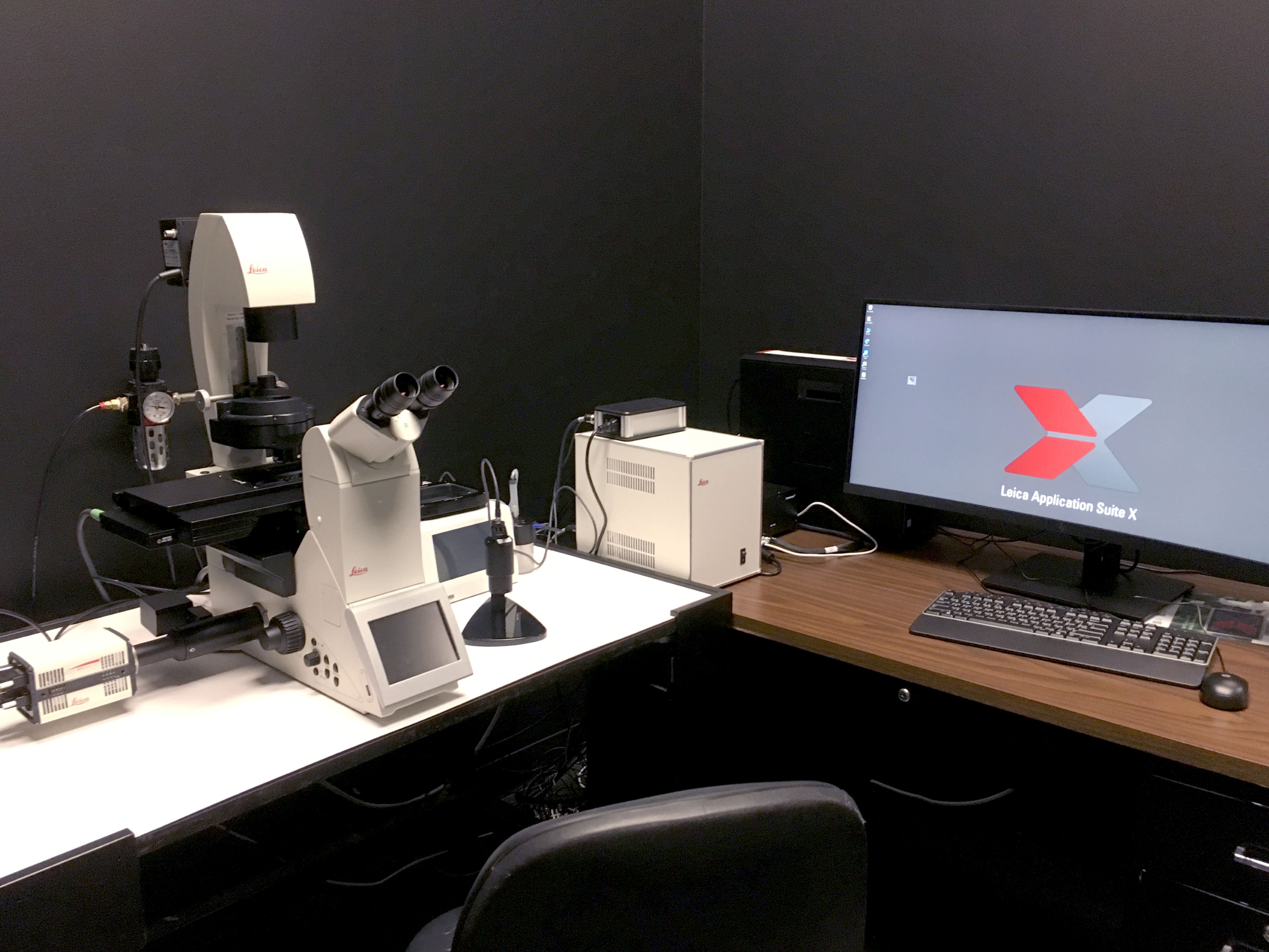
Leica Stellaris Confocal Microscope
Equipped with four laser lines(405nm, 488nm, 561nm, and 638nm). The four HyD S detectors provide the ability to separate fluorochrome signals with overlapping emission characteristics and thus further increases flexibility for studies involving multiple fluorescent labels. The system is mounted on a Leica DMi8 inverted microscope with a motorized stage.
Applications that can be performed on this machine include: XY images, Z-stacks, time series, bleaching experiments (including FRET and FRAP), and multiple position imaging (including tile scans).
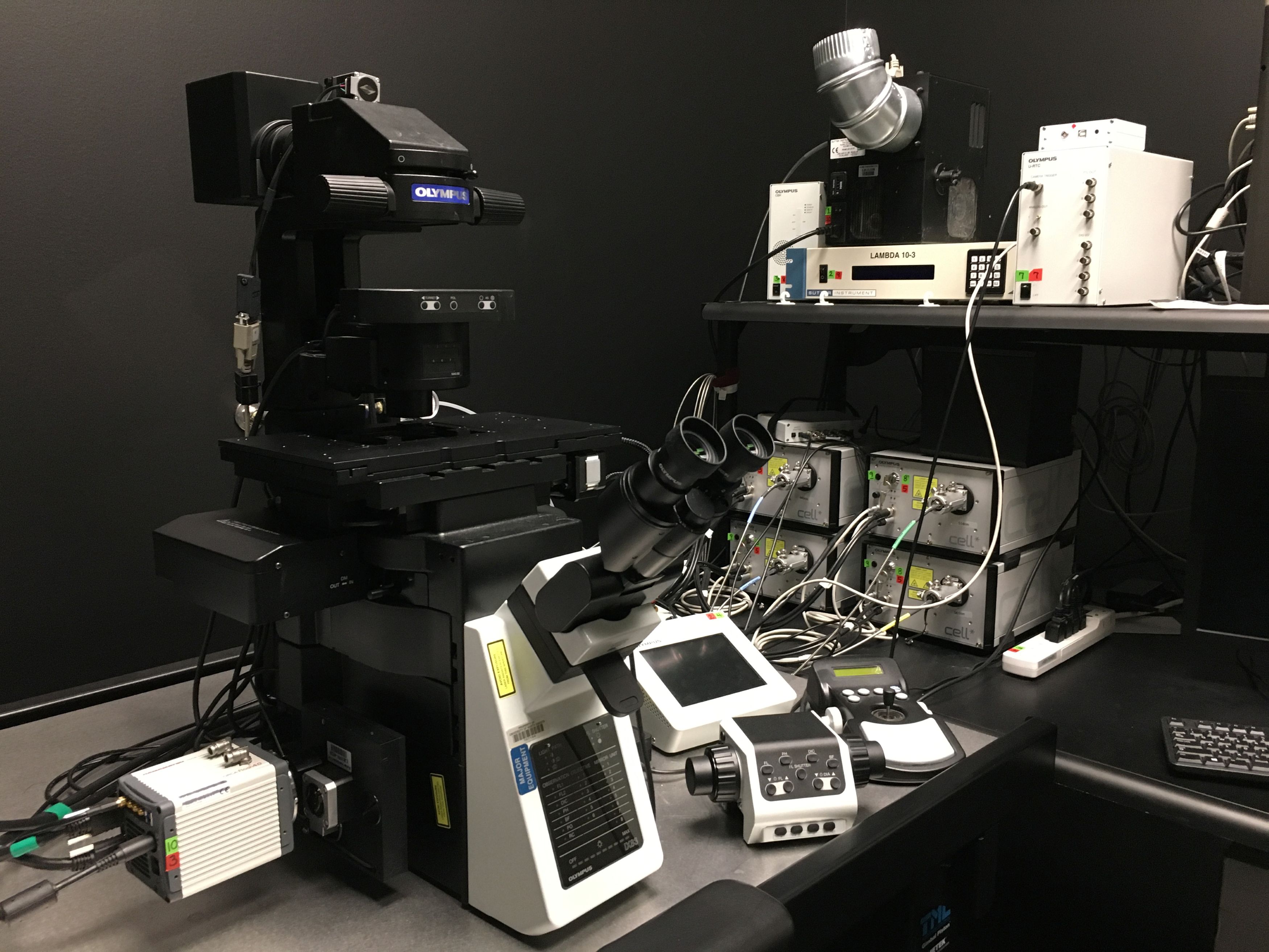
Olympus FV1000 MPE Basic Multiphoton Microscope
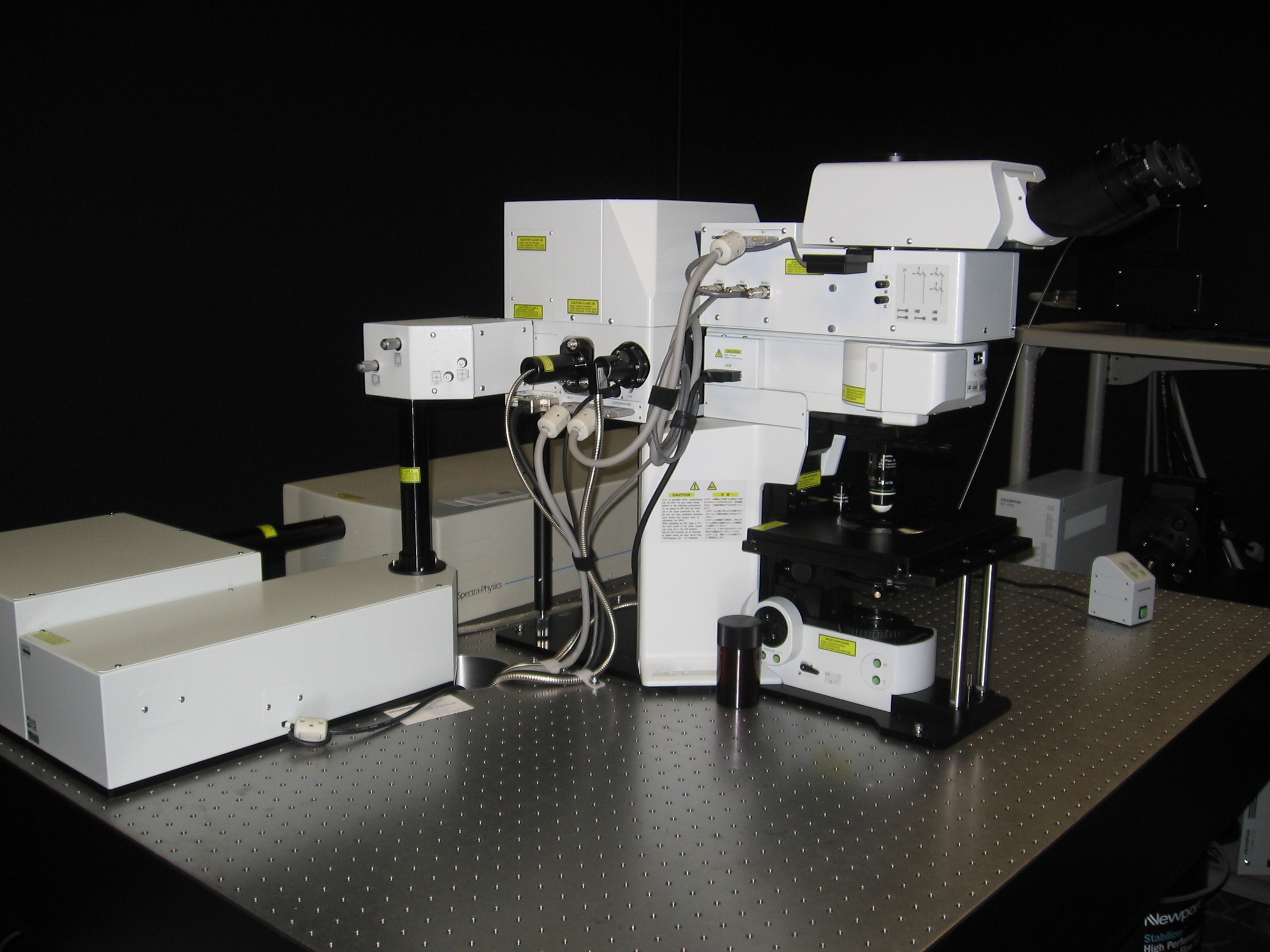
The system is mounted on an Olympus BX61WI upright microscope and equipped with a mode locked Ti:sapphire laser (690-1040nm), a multi Argon laser (457nm, 488nm, 515nm, Total 30mW), a HeNe laser (543nm, 1mW), and a 635nm HeNe laser. The system is configured for a wide range of applications: single photon imaging, multiphoton imaging, XY imaging, Z-stacks, time series, bleaching, and intravital microscopy using small animals.
Brightfield/Fluorescence
Nikon Eclipse 80i (upright) Fluorescent Microscope
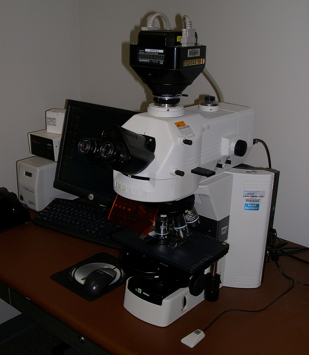
Equipped with a SPOT II digital camera capable of capturing black and white as well as color images. Emission filters are available for DAPI, FITC, Rhodamine, and Texas Red.
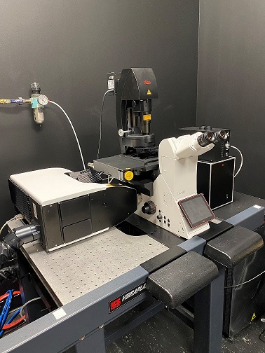
.Leica Thunder Imager
Mounted on an inverted DMi8 microscope the Leica Thunder Imager is also equipped with a high resolution CMOS camera (up to ~1000fps) and a high resolution software controlled stage. LED light sources are tuned to 395, 440, 470, 510, 550, and 640nm. For live cell imaging there is a stage top incubator with CO2. Instant computational clearing can be done on single plane images or you can perform iterative 3D deconvolution by taking multiple plane images (z-stacks) and letting the system apply the algorithm as you scan.
TIRF Systems
Olympus TIRF Microscope
Equipped with a xenon light source as well as 405nm, 491nm, 514nm, and 561nm lasers. It can perform both widefield fluorescence and TIRF applications. Two different cameras are available; a Hammamatsu RCA flash 4.0 camera or an Andor iXon Ultra camera. The system is mounted on an inverted Olympus IX83 microscope with an automated stage.
Applications that can be performed include: XY imaging, Z-stacks, time series (including streaming), calcium imaging, and multiple stage position imaging. Live cell imaging of round dishes is supported with a Tokai Hit stage heater and CO2 delivery system.
Auxiliary Equipment
- Attofluor Metal Chambers (6 qty) that hold 25mm round coverslips
- Harvard Apparatus Temperature controller; model TC-202A
- Medical Systems Corp: Open perfusion Micro-incubator; model PDMI-2
- Sanyo CO2 Incubator; model MCO-5AC
- Refrigerator
- Stage Top Heaters: These are interchangeable on any of our inverted systems.
- Okolab: Can accommodate plates, 35mm dishes, and chamber slides
- LCI: Can accommodate plates, dishes (35mm, 50/60mm), and chamber slides
- Tokai Hit: Single 35mm dish capacity
Imaging Core Workstation Computer (Stand Alone)
- Equipped with the following software
- FIJI - Multi purpose software used to open a multitude of file types and perform varies types of analysis
- Huygens Essential - Deconvolution software
- Imaris (v 9.3.0) - Analysis software
- Includes the Imaris for Tracking package.
- Leica Application Suite X (LAS X) - Offline application support for Leica files