Teaching Files
MRI - Shoulder Anatomy
-
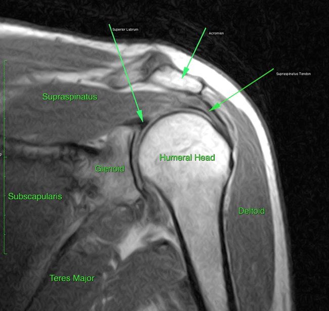
Shoulder 1
-
Shoulder 2
-
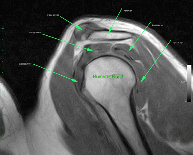
Shoulder 3

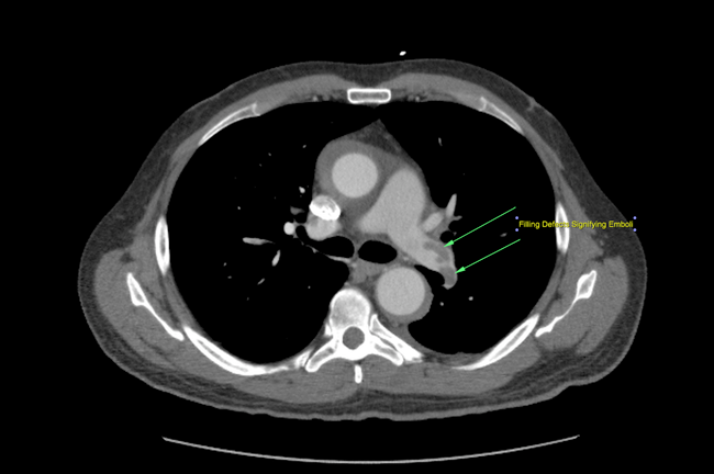
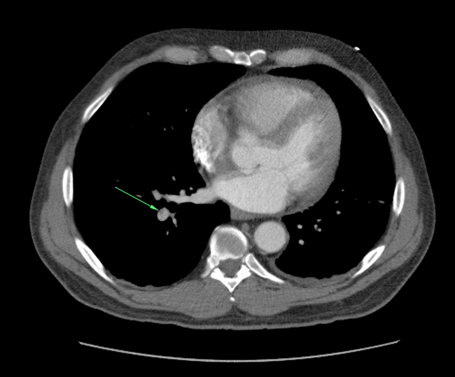
Bilateral Pulmonary Emboli
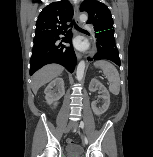
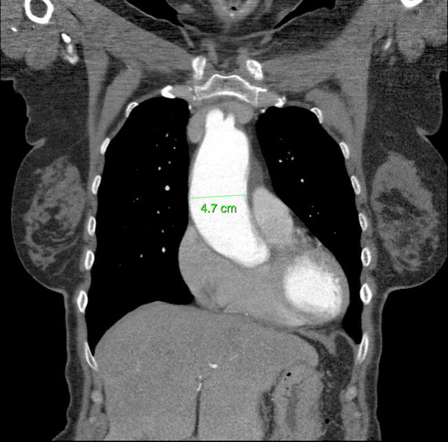
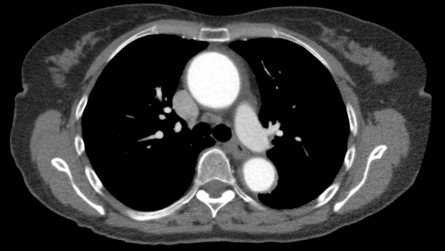
Ascending Aortic Aneurysm
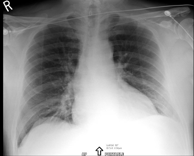
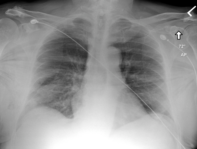
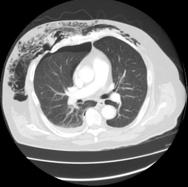
Subcutaneous Emphysema
-
Notice muscle fibers of pectoralis major muscles indicating air outside of the pleura
-
This finding is highly suggestive of pneumothorax (although not obviously seen on this plain film)
Follow-up CT was obtained
-
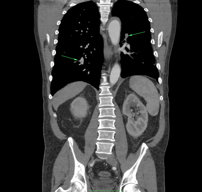
Pneumothorax noted to right lung along with significant subcutaneous emphysema
-
Also note fracture of rib on the right
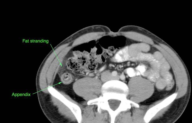
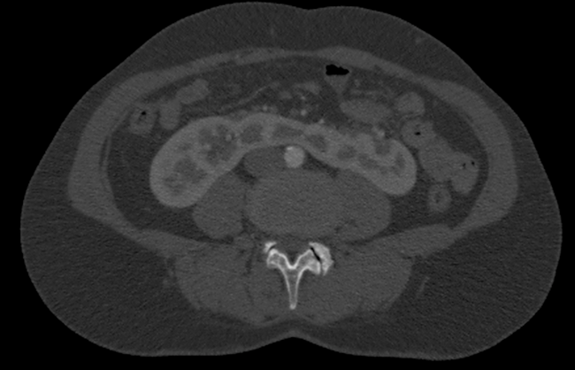
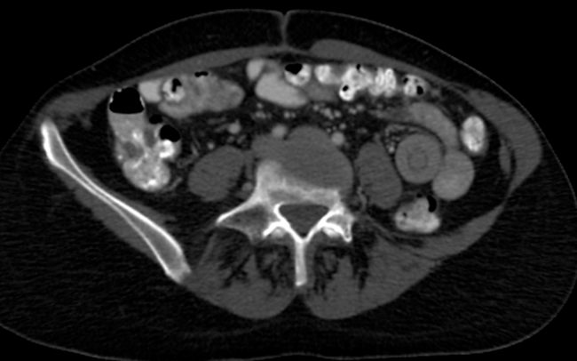
Appendicitis
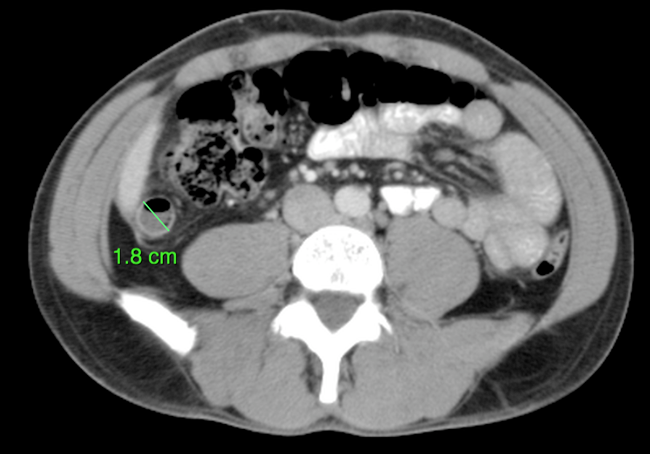
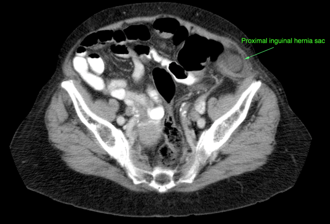
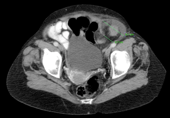
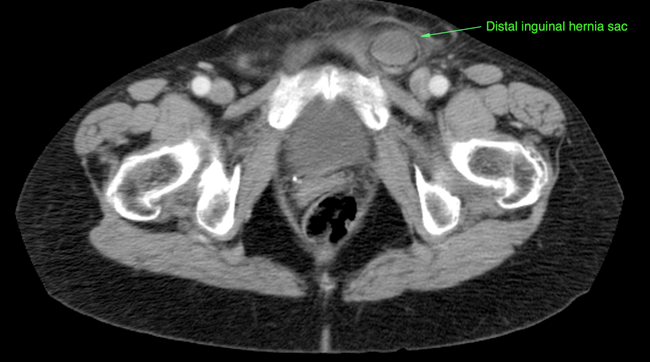
Incarcerated Inguinal Hernia
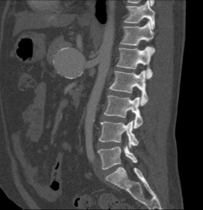
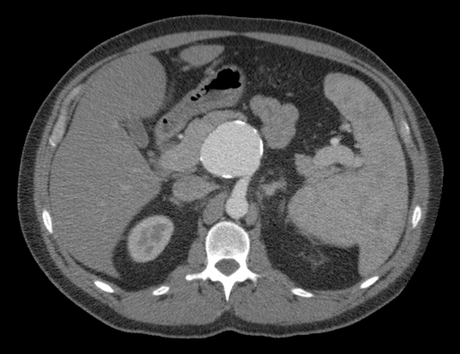
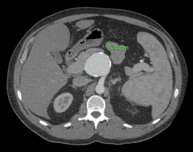
Superior Mesenteric Artery Aneurysm
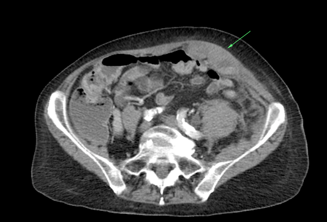
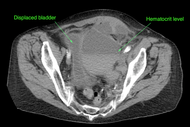
Rectus Abdominis Hematoma
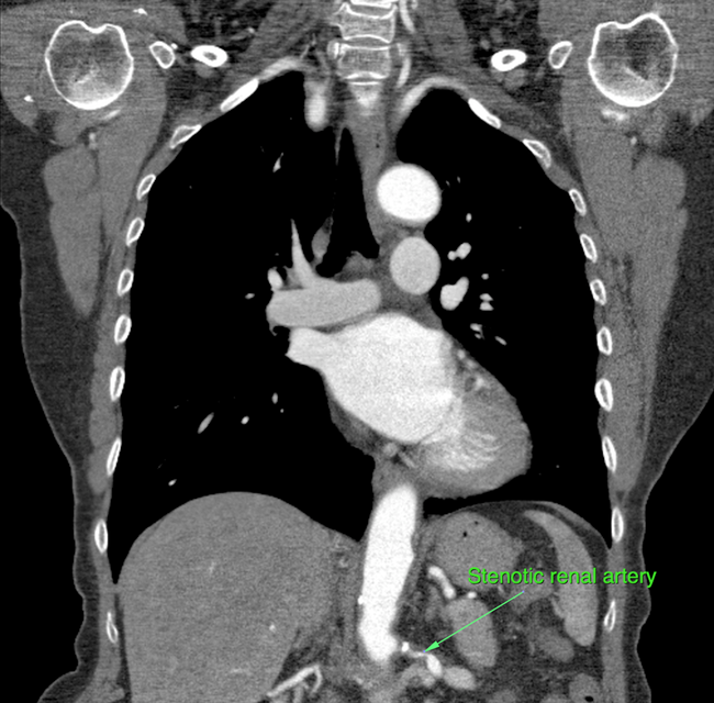
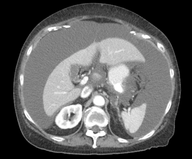
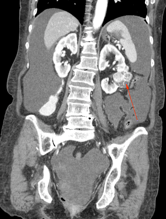
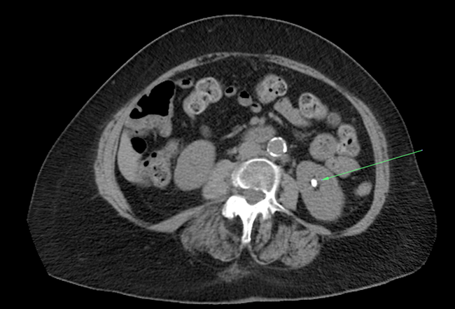
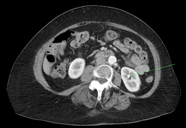
Renal Artery Stenosis
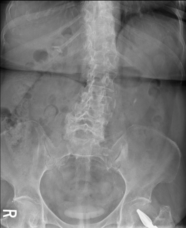
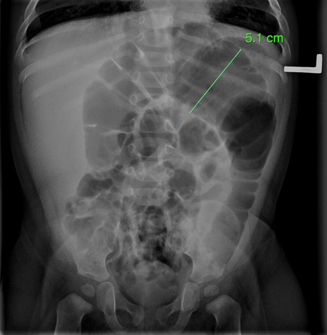
Small Bowel Obstruction
-
Multiple air-fluid levels
-
No gas in rectum
-
Dilated loops of small bowel
-
Plicae circulares seen across entire diameter of small bowel.
Ascites
-
Note the renal mass. Since it is not cystic in appearance it is malignancy until proven otherwise.
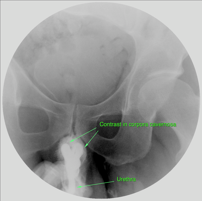
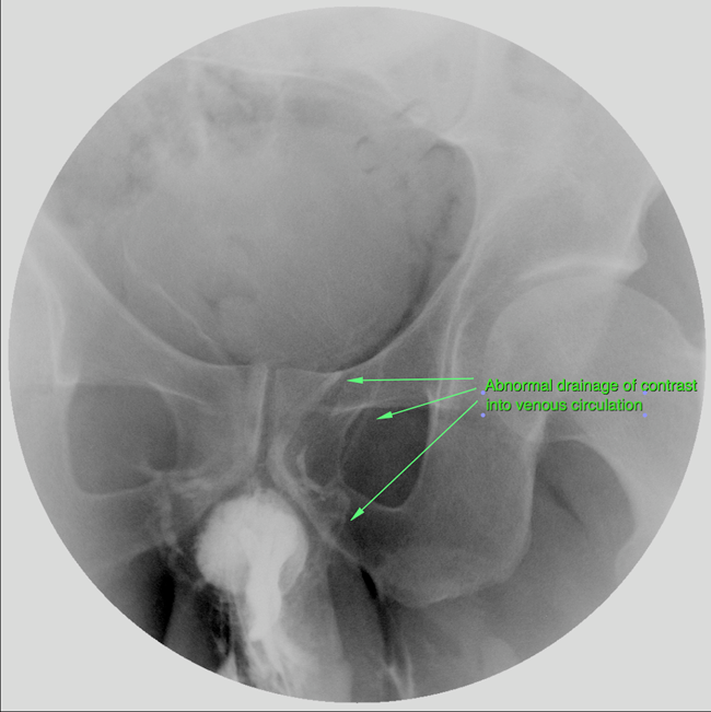
Urethral Trauma (Male)
Horseshoe Kidney

Nephrolithiasis
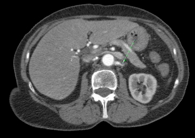
Lobar Nephronia
-
Low density mass with ill-defined borders in left kidney
-
Due to infection from vesicoureteric reflux
-
DDx - Abscess
-
Tx - IV antibiotics
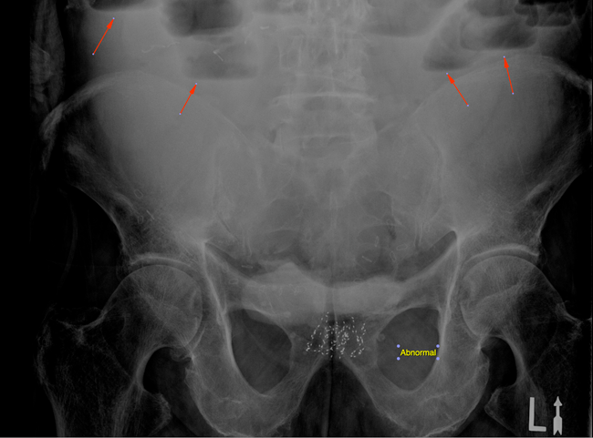
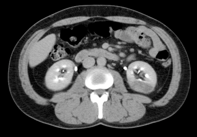
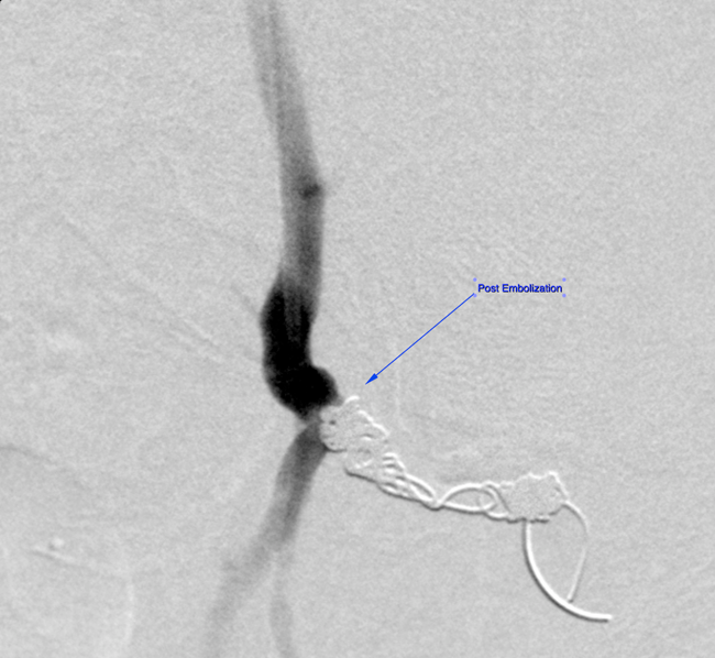
Superior Gluteal Artery Coil Embolization Pos-Pelvic Trauma
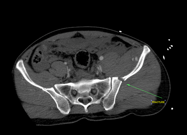
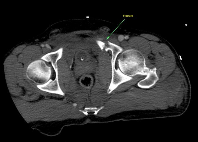
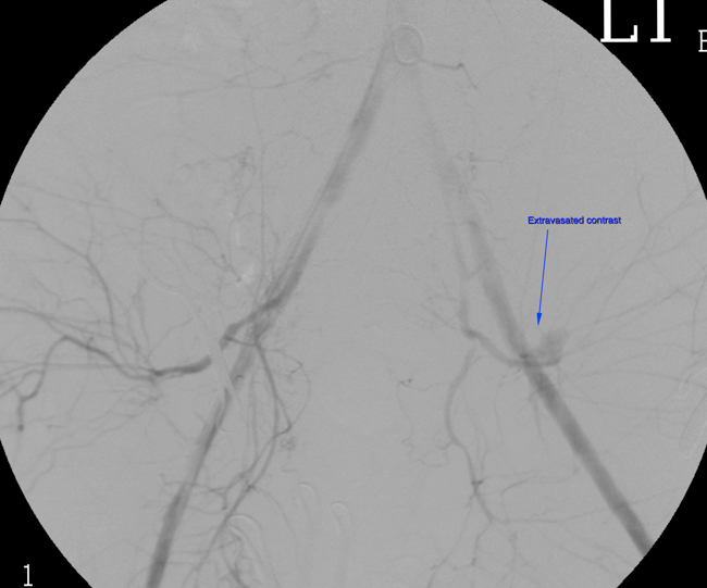
The above still image, taken from the angiography movie below, demonstrates contrast material extravasating from the superior gluteal artery. This artery was lacerated during the traumatic event, an automobile accident, that produced the fractures of the pelvic bones seen in the above CT images. The movie then shows the coil device within the lacerated vessel, and injection of contrast material allows visualization of the successful embolization of the superior gluteal artery.
- This photo shows the deployment of the inferior vena cava filter this patient received.
Breast Cancer
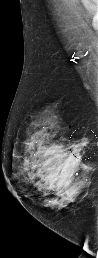
70 yo woman with history of cancer in the right breast presents for screening mammography as shown below.
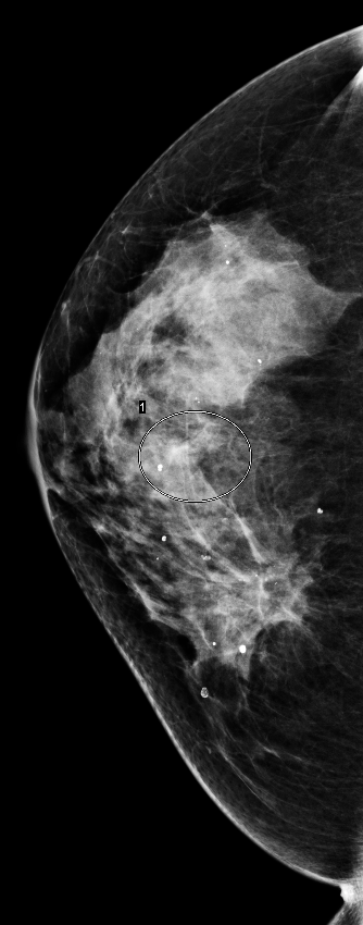
- There is a suspicious density noted in the mid-portion of the right breast.
- Surgical clips are noted from prior intervention in the right breast.
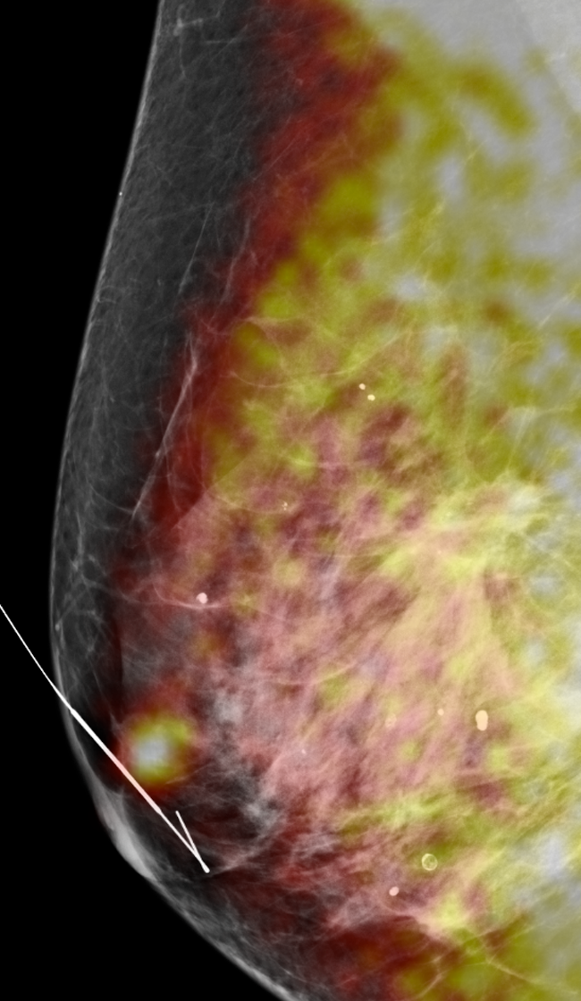
- BSGI is performed and shows a hot nodule just deep to the nipple of the right breast. Note that the lesion seen on mammo does not enhance on BSGI. The nodule found on BSGI was not seen on the standard screening mammo.

- The patient was taken for mammographic-guided needle placement for surgical removal. The color enhancement of the next following image is an overlay of the BSGI data on the mammographic data done post needle insertion to assure correct placement of the needle for surgical intervention.
- Breast tissue s/p surgical removal. Pathologic exam found this to me a second primary breast cancer in this 70 yo.
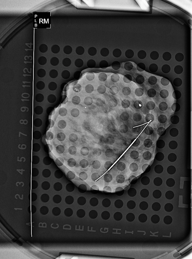
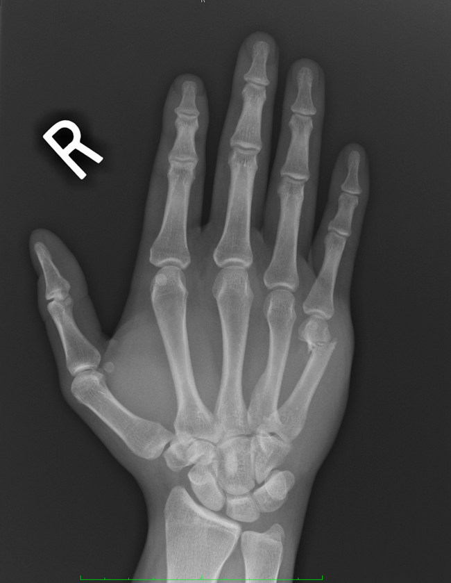
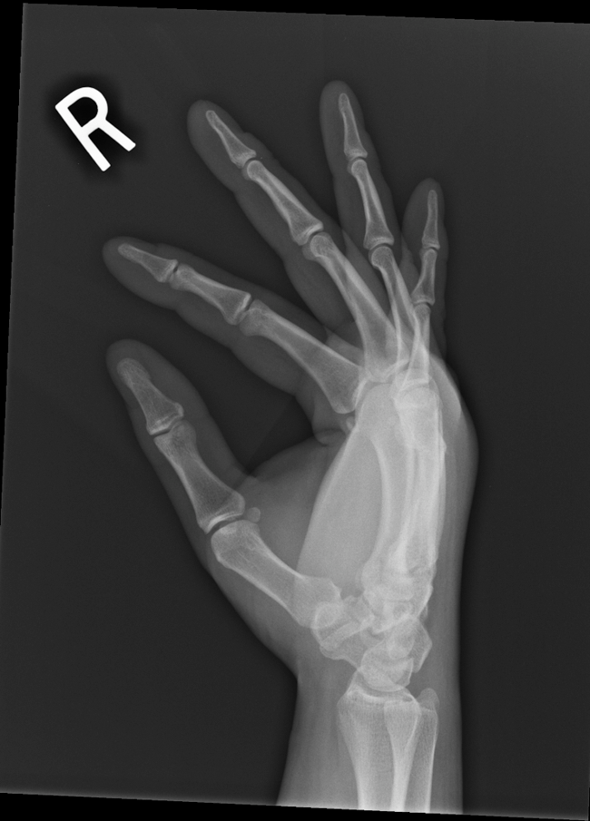
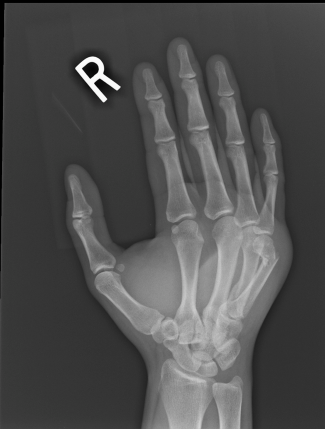
Boxer's Fracture
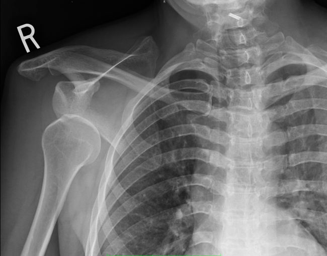
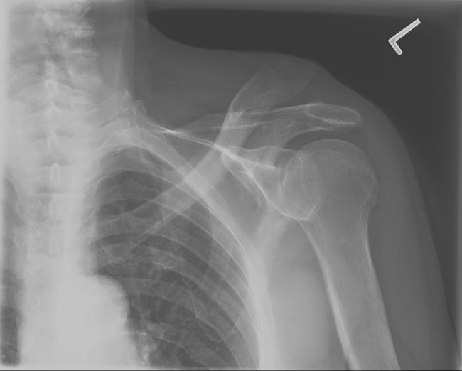
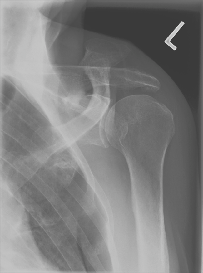
Anterior Shoulder Dislocation
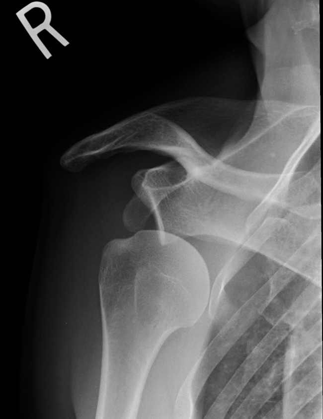
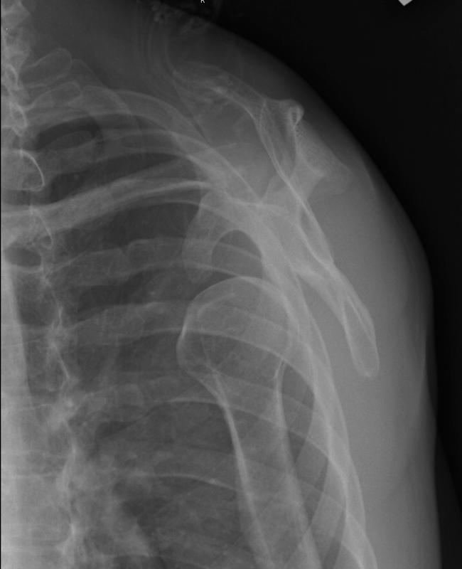
Chronic AC Joint Separation
Subarachnoid Hemorrhage
Encephalitis
Ischemic Stroke With Hemorrhagic Conversion
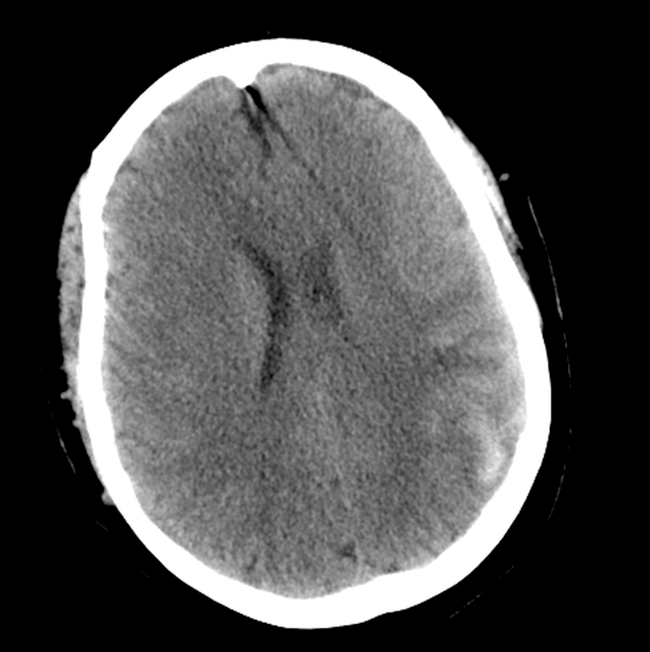
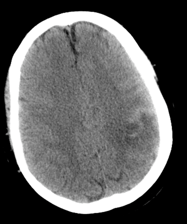
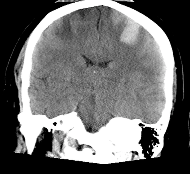
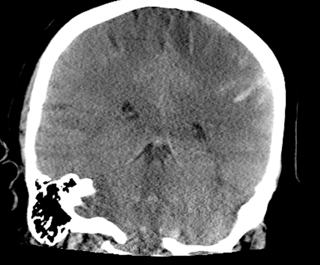
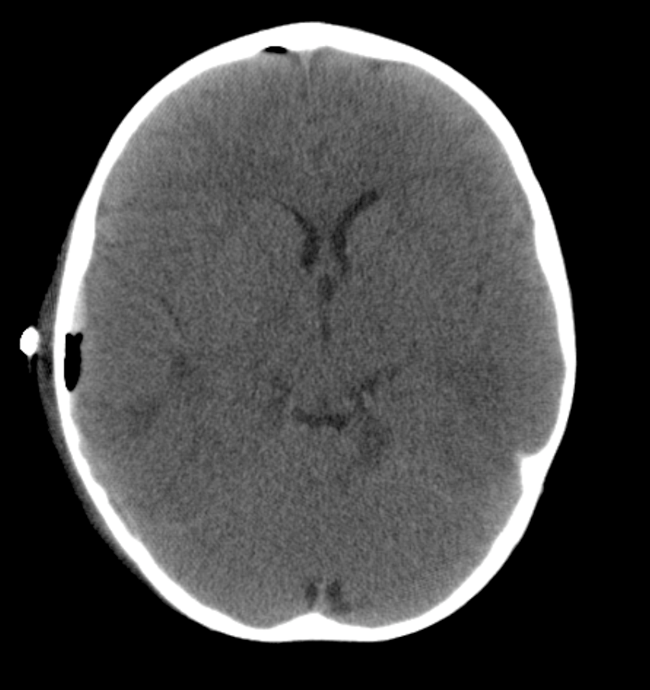
Epidural Hematoma (Pediatric)

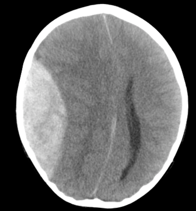
These images were taken of a patient who had become unresponsive approximately 10 hours after falling from a deck. These coronal and axial CT images (above) show an obvious epidural bleed demonstrating the classic lentiform shape and mass-effect with a shift in the midline structures. However, there is no apparent temporal fracture as would be expected with such a finding. A craniotomy was preformed for decompression and a subsequent CT (below) shows resolution of the mass effect.
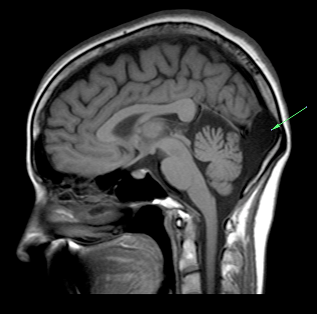
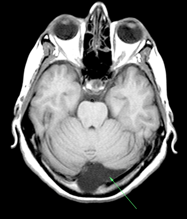
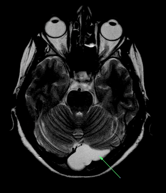
Arachnoid Cyst
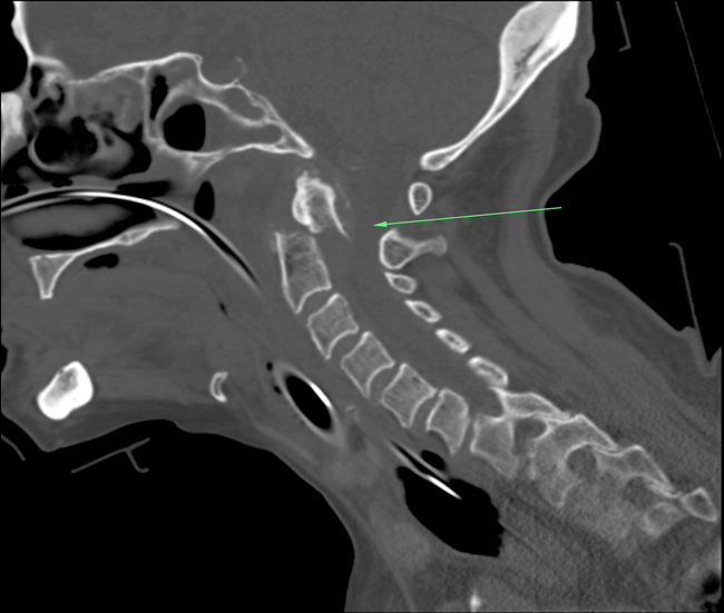
Odontoid Fracture
Hirschsprung Disease
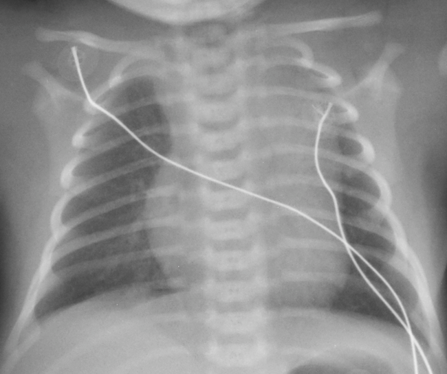
Pneumothorax (Newborn)
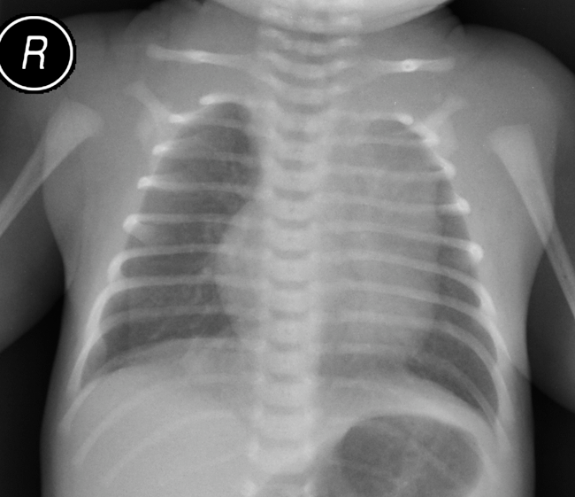
This is a frontal chest radiograph (above) taken of a newborn with shortness of breath. The density in the left upper thorax is actually a normal thymic shadow. In the right lower lung field there is what appears to be a pneumothorax, which, with respiratory treatment, has resolved on the subsequent radiograph taken the next morning (below).
Obstipation
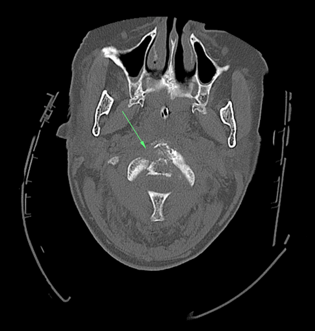
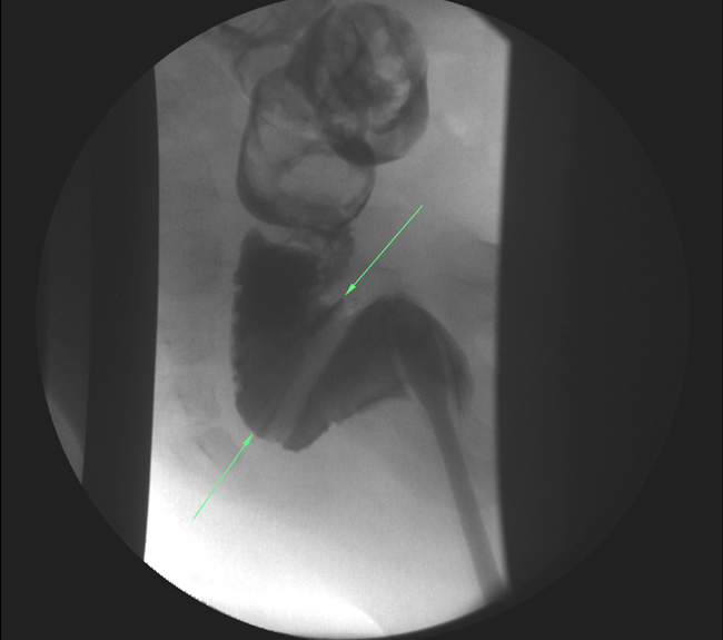
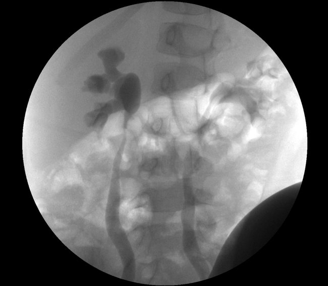
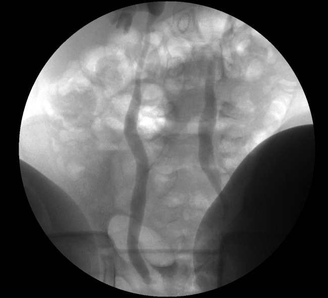
Vesicoureteral Reflux
The video link above demonstrates a series of still images obtained during a voiding cystourethrogram. The still images below display marked dilatation of the ureter and calyces suggesting grade 4 reflux.
Pyloric Stenosis
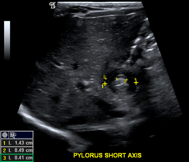
3-7-15 Rule:
- < 3mm single muscle thickness
- < 7mm pyloric width
- < 15mm pyloric length
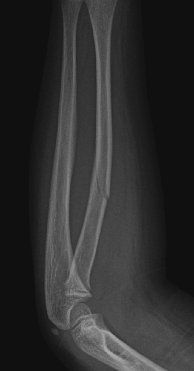
Greenstick Fracture
Intussusception
Adult Etiology
- Cancer most likely source of lead point
Pediatric Etiology
- Idiopathic
- Meckel's Diverticulum
- HSP
Pediatric Signs & Symptoms
- 3mo to 6yo (most common 7-8mo)
- Colicky abdominal pain
- Currant jelly stool
- Palpable sausage-like mass
Air or contrast enema is both diagnostic and therapeutic.